
 3405 8288
Olympus EVIS X1 endoscopic series
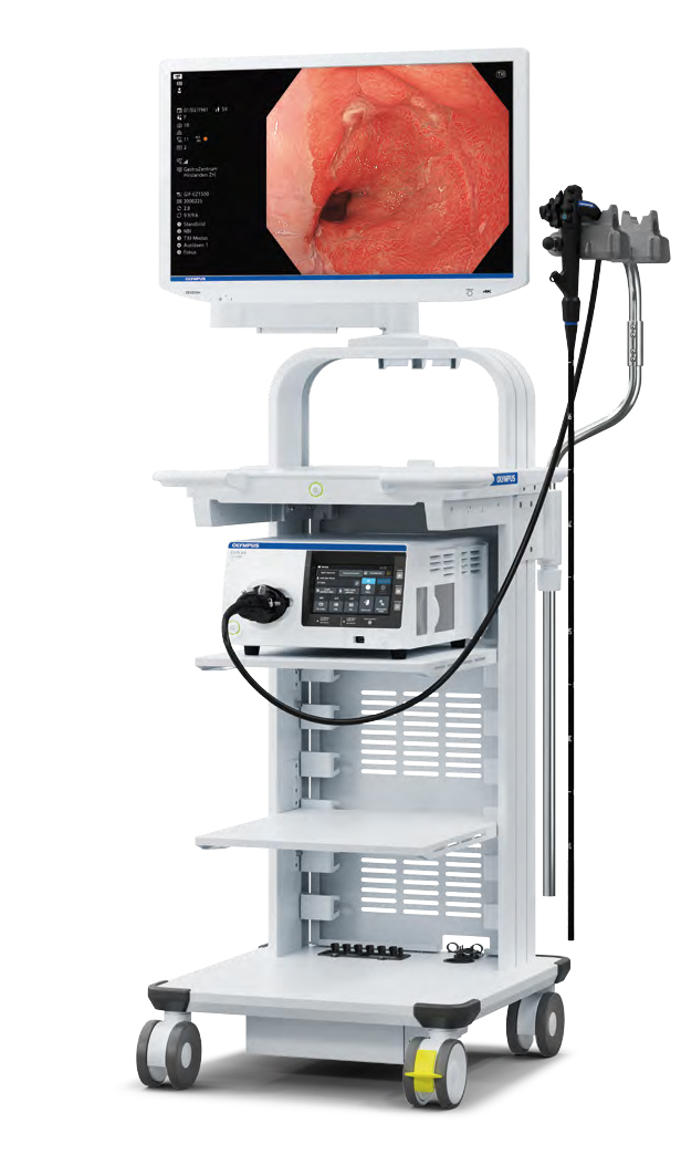 |
Since 2020, The Specialists have employed the Olympus EVIS X1, a state-of-the-art endoscopy unit enhanced with artificial intelligence. This advanced technology offers superior imaging capabilities, including:
The integration of AI with these advanced imaging features significantly improves the detection and diagnosis of polyps and pre-cancerous lesions, ensuring high-quality patient care.
Recent research indicates that the use of Artificial Intelligence in colonoscopy significantly improves the rates of polyp and adenoma detection, irrespective of a patient's medical history, family background, sex, or lifestyle habits. At The Specialists, we are dedicated to providing the highest standard of care by remaining at the forefront of innovative technology. |
 |
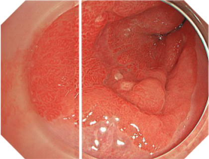
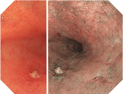
Texture and Color Enhancement Imaging, TXI
TXI technology aims to enhance the visibility of potentially suspicious tissue, using a white-light imaging effect that improves the colour, structure and brightness. By supporting better visibility of potential lesions, TXI aims to contribute to higher detection rates.
|
|
|
 |
Extended Depth of Field, EDOF
EDOF allows precise observations through continuous broad focus and seamless magnification. This continuously sharp image is developed to reduce the necessity for focal adjustments and to even contribute to easier identification and a more confident abnormality diagnosis. |
|
|
 |
5-Led Spectrum Technology
The EVIS X1 video system centre contains five LEDs that are combined to produce different observation modes. It includes an amber LED tailored by Olympus, enabling the visualization capabilities of the RDI mode. |
 |
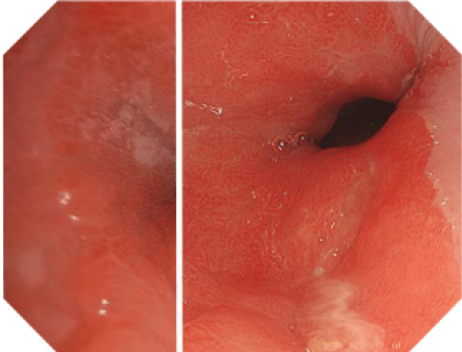
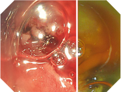
Red Dichromatic Imaging, RDI
RDI is designed to enhance the visibility of deep blood vessels and bleeding sources. Easier identification of bleeding spots makes haemostasis quicker and easier. This helps to reduce the stress of the physician during endoscopic therapy. |
|
|
 |
Computer-Aided Diagnosis, CAD
The next upcoming feature of the EVIS X1 system will be computer-aided diagnosis (CAD) using artificial intelligence to integrate breakthroughs in deep learning into the world of image detection, characterization and staging. |
 |
Narrow Band Imaging, NBI
Utilizing specific blue and green wavelengths absorbed by haemoglobin, NBI creates a strong contrast between vessels and surrounding mucosa.11 This facilitates the visibility of highly vascularized areas, blood vessel patterns and surface structures that are predictive for distinct histopathologies. |
|
|
 |
Dual Focus– Two-Stage Optical Lens Technology
Allows switching from normal focus to near focus mode with a single button, to conduct close examination of mucosal tissue and capillary networks. |
 |
4K Monitor and New Symbol Indication
4K UHD LCD Monitor optimized for Olympus Endoscopy Systems and 4K Upscaling for conventional HD imaging systems. |
 |
Responsive Insertion Technology, RIT
Combines PB (Passive Bending), HFT (High Force Transmission) and variable stiffness to improve ease of insertion and operator control. |
 |
Brightness Adjustment Imaging with Maintenance of Contrast
BAI-MAC (Brightness Adjustment Imaging with Maintenance of Contrast) is a new image processing technique to improve the brightness levels in dark areas of the endoscopic image, whilst maintaining the brightness of lighter areas to increase the distance view. |
 |
Water Jet
Improves the accuracy of observations and efficiency of treatment by easily removing mucus and other residues in the investigated intestinal areas. |